|
COMPUTERIZED ANATOMY ABSTRACT The segmented image information is stored in two independent files. A variable size file is created for each transverse slice and contains the x,y coordinates of each of the contours drawn on that slice. The slice number is retained in the name of the file. DEPARTMENT OF DIAGNOSTIC RADIOLOGY |
BRAIN Computerized3-DimensionalSegmentedHumanAnatomyI.GeorgeZubal,Char SKULL nalphantomvolume.Thecentralvoxel'sfilteredvaluewascalculatedasth heavailableUserInterfaceServices(UISroutines)forprogramcontrolof mforECT.'J.Nucl.Med.,27,1577-85,(1986).[12]CEFloyd,RJJaszczak,KL -123.'Radiology,164,279-81,(1987).[13]JWBeck,RJJaszczak,REColema nNuclearScience,29,506-511,(1982).[14]DAcchiappati,NCerullo,RGuz EdColchesterandHawkes,8-22,(1991).LEGENDSTOFIGURESTABLE1:Listofo eousphantom.NM/MIRDPamphletNo.5.,(SocietyofNuclearMedicinePublic 1-407,(1992).[17]RMohan,GMageras,BBaldwin,LBrewster,GKutcher,SLe lstothedisplayedtransverseimages.AseriallinehighresolutionSummag orrespondingtotheorganindexvalue.Theassignmentofintegerstotheorg redondiskwitharesolutionof512by512pixels.Membersofthemedicalstaf therapy.'Med.Phys.,18,43-53,(1991).[19]IGZubal,CHHarell.'Voxelba SPINE 'AScatterCorrectionMethodforT1201Images:AMonteCarloInvestigation exsliceimagescanbereadintoa512by512by119voxel3-dimensionalarray, ages.'Phys.Med.Bio.,30,239-49,(1985).[7]DRDance,GJDay:'Thecomput inSPECT.'J.Nucl.Med.,26,403-8,(1985).[11]CEFloyd,RJJaszczak,KLGr nformaltreatments.'Med.Phys.,19,933-944,(1992).[18]DNigg,PRandol 5).[6]HKanamori,NNakamori,KInone:'EffectsofScatteredx-raysonCTim onalmatrices(eachcollapsedmatrix=128x246)byaddingallintegervalue Brain20-Pancreas3-Spinalchord21-Adrenals4-Skull22-Fat5-Spine23-B TWehavemanuallysegmented129x-rayCTtransverseslicesofalivingmaleh onte-CarloSimulationTechniques.'EuropeanJournalofNuclearMedicine -Lymphnodes10-Lungs28-Thyroid11-Heart29-Trachea12-Liver30-Diaphr .[20]IGZubal,CRHarrell,PDEsser.'MonteCarlodeterminationofemergin SPINAL CHORD ferenceonInformationProcessinginMedicalImaging,,Springer-Verlag, yYaleUniversitySchoolofMedicine,NewHaven,CT06510correspondingaut n,CFStarmer,LWNolte.'AnalysisofSPECTIncludingScatterandAttenuati ion,BenjaminM.Tsui,Ph.D.,Dept.ofRadiology,Univ.ofNorthCarolina.2 gr.,7,316-20,(1983).[9]CEFloyd,RSJaszczak,CCHarris,REColeman:'En ,15,683-686,(1989).[15]MSaxner,ATrepp,AAhnesjo:'Apencilbeammodel toindexnumbers:4,5,6,7,and8).FIGURE2:Anteriorandlateralprojectio jorinternalstructuresofthebody.Eachvoxelofthevolumecontainsanind esolutionof1millimeterinthex,yplane.Thez-axisresolutionis1centim uter-basedmodelingandsimulationcalculations.keywords:segmentatio mscaneitherbedefinedbymathematical(analytical)functions,ordigita frominternalorexternalsourcesofradioactivityandservedtocalculate ADRENALS herethestructuresadjacenttotheheartandtheheartitselfhavebeenmore rticularlyforanalyzingscatterandattenuationproblemsinnuclearmedi 5-18].Thesenewtherapytechniquescanbemoreeffectivelyinvestigatedw onsiderablenumberofpatientsareimagedfromheadtomid-thighinourhosp ltlesionsofapproximately0.5,1.0,and2.0centimeterdiameterwerearbi -dimensionalvoxelphantomofthehuman.SincetheoriginalCTimagesarest rdisplay.DISCUSSIONWehavecreatedadigitalvoxel-basedphantomwhichc hnotonlycloselyresembleclinicaldata,butincludeadditionalinformat gthisveryrealistichumanmodelisthatsuchsimulationscandecreasethen dRealisticHumanPhantomsandMonteCarloMethods',Phys.Med.Bio.,31,44 eer,REColeman:'InverseMonteCarloasaUnifiedReconstructionAlgorith sedMonteCarloCalculationsofNuclearMedicineImagesandAppliedVarian LYMPH NODES rganindexnumbersandrespectiveorgansorstructuressegmentedinthehum am13-Gallbladder31-Spleen14-Leftandrightkidney32-Urine15-Bladder Inordertogainaccesstothephantomdata,pleasesendyourrequesttoDr.Ge canthenbeindexedtoactivitydistributionsorotherphysicalcharacteri 4pixelrasterdisplayequippedwith8bitplanes.Onebitplaneisusedforov sforPhotonSourcesUniformlyDistributedinVariousOrgansofaHeterogen romtheCTarchivereeltoreelmagnetictape,decompressingtheimagesfrom 8x246matrixwheretheisotropiccubicvoxelresolutionis4millimeterson orpublicaccessthroughourImagingScienceResearchLaboratory.2RESULT salongrowsofvoxels.Thefinalmatriceswerenormalizedanddisplayedusi ndtherapy.Wehopetoextendtheapplicationofthisphantomtotherapyrela zardi:'AssessmentoftheScatterFractionEvaluationMethodologyusingM THYROID gooni,RNath:'Tissueinhomogeneitycorrectionforbrachytherapysource ibel,CBurman,C.Long,ZFuks:'Clinicallyrelevantoptimizationof3-Dco ett,HBBarber,JMWoolfenden.'SurgicalProbeDesignforacoincidenceIma gingSystemWithoutaCollimator'Proceedingofthe12thInternationalCon fDiagnosticRadiology,BML332333CedarStreetNewHaven,CT06510ABSTRAC ansareshowninTable1.DataAccessandDisplayThe2-dimensionalorganind dertomakethedatamoremanageableandtohaveconsistentvoxeldimensions eachside.Thisvolumearrayiscreatedbycombiningpixelsinthex,yplanea mantorso.Suchananthropomorphic3-dimensionalphantomhasseveralinte ingthesegmenteddata.WeareindebtedtoCorneliusN.deGraafUtrecht,The ObjectModelforMonteCarloSimulatedEmissionandTransmissionTomograp MonteCarloinvestigation.'Phys.Med.Bio.,29,1217-30,(1984).[10]CEF TRACHEA onUsingSophisticatedMonteCarloModelingMethods.'IEEETransactionso lsegmentedhumanphantom.Theskinandfat(indexnumber=1)arehighlighte ngs(10),bladderandureters(15),andbone(34).FIGURE3:Anteriorandlat ImagingScienceResearchLaboratoriesDepartmentofDiagnosticRadiolog umanandcreatedacomputerized3-dimensionalvolumearraymodelingallma fallowingveryfastcalculationoftheintersectionofraylineswiththean alyticalsurfaceswhichdelineatetheorgans.Aversionofthismathematic allyversions1ofthephantomwhichareusedfordedicatedcardiacstudiesw runderstandtheimageformationprocessindiagnosticradiology[4-7],pa cdistributionscannotbeadequatelyevaluatedbysuchsimplegeometries. Inthefieldofoncology,internalandexternalradiotherapysourceshaveb dx-raytomography(CT)suppliesustherequiredhighresolution3-dimensi DIAPHRAM italtostudydiffusediseases.Weselectedanadultmalewhosedimensionsw of78sliceimageswereacquiredfromnecktomid-thighwitha1centimetersl icethicknessusinga48centimeterfieldofview(pixelsize=1mm).Duringa theresidentcolordisplayscreen.Thecolordisplaymonitorisa1024by102 amwasdevelopedtoreadthetransverseslicesfromdisk,displaythemonthe sandcontoursweredrawnwith1millimeterresolutiontodefinethisfully3 linthedefinedstructures.Thescannerusedisaclinicalinstrument;thea transversesliceinwhichtheHounsfieldnumbersarereplacedbyintegersc inwhichthex,yresolutionis1millimeterperpixelandtheslicethickness ctedanteriorandlateralviewsofselectedstructuresfromthe128x128x24 ntegervalueandsettingother(unselected)structuresto0.The3-dimensi onalvolumewasthencollapsedparalleltothemajoraxesontotwo2-dimensi RIB CAGE hepatient'sbody;withinthisoutline,variousstructuresareselectedfo ionnotdeterminableinpatientstudies.Suchdatasetscanhelptobetterun canproveespeciallyinterestingintestingandimprovingtomographicrec g'invivo'simulations.Thetumordetectioncapabilitiesofanovelcoinci ansmissiondiagnosticradiology.'Med.Phys.,16,:490-5,(1986).[5]HPC ph,FWheeler:'Demonstrationofthree-dimensionaldeterministicradiat genergyspectrafordiagnosticallyrealisticradiopharmaceuticaldistr ibutions.'NuclearInstrumentsandMethodsinPhysicsResearch,A299,544 anphantom.FIGURE1:Anteriorandlateralprojectionsofthe3-dimensiona dtoshowtheoutlineofthepatientaswellasinternalbones(corresponding alphantomhasbeenupdatedtoincludefemaleorgans[2].Thereareaddition themanufacturer'slosslessstorageformat,andstoringtheminexpandedm SKELETAL MUSCLE 129to119slicesisduetotheoverlapofinformationintheneckregion.Inor nwithmillimeterresolutionineachof129transversesliceimagesofthehu derstandtheimageformationprocessforclinicallyrealisticmodels,and esandattenuatingmedia.Suchsimplegeometriesareusefulinstudyingmor ntainedinthetransverseslices.Aregionofinterestcoloringroutinewas onstructionalgorithms[21].Newimagingdevicescanbeinvestigatedusin Greer,REColeman:'BrainPhantom:highresolutionimagingwithSPECTandI 33-Feces(coloncontents)16-Esophagus34-Testes17-Stomach35-Prostat secondimagingsession,51slicesofthesamepatientwereacquiredofthehe oourimageprocessinglabbyreadingthereconstructedtransverseslicesf SInordertoappreciatethedetailoftheanthropomorphicphantom,weproje orphicphantomswasdevelopedforestimatingdosestovarioushumanorgans LUNGS linders,orrectangularvolumes.Forinternaldosimetrypurposes,suchhu ecomemoresophisticatedintheirdesignandapplications.Thecalculatio nsinvolvedinclinicaltherapyplanninghavebecomemoresophisticated[1 stics(densityorelementalcomposition).Wehaveconstructedananatomic adandneckregionwith5millimeterslicethicknessandafieldofviewof24c erlaygraphicswhiletheremaining7bitsareusedformapping128colorleve raphicsbitpadprovidedhighresolutioncursorcontrol.Anin-houseprogr edforeachtransversesliceandcontainsthex,ycoordinatesofeachofthec is10millimetersinthebodyand5millimeterinthehead.Thereductionfrom ndbyduplicatingslicesalongthez-direction.Inordertoremovesomeofth ontours=1Megabyte,organindexmatrices20Megabytes,andareavailablef restingapplicationsintheradiologicalsciences.Wehaveroutinelyused HEART CES[l]WSnyder,MRFord,GWarner:EstimatesofSpecificAbsorbedFraction ergyandspatialdistributionofmultipleorderComptonscatterinSPECT.A ceReductionTechniques.'ImageandVisionComputing,10,342-348,(1992) eralprojectionshighlightingtheskinandfat(indexnumber=1),andmyoca 9-452,(1986).[3]H.Wang,RJJaszczak,REColeman:'SolidGeometry-Based hehumananatomyserveanimportantroleinseveralaspectsofdiagnostican theS-factorsforinternaldosecalculationsinnuclearmedicine[1].This mathematicalphantommodelsinternalstructuresaseitherellipsoids,cy nshavesometimesbeenlimitedtosimplepoint,rod,andslabshapesofsourc illavailable,theoriginalHounsfieldnumbersarealsoknownforeachvoxe ceandcalibrationcarriedoutforqualityassurance.Thesegmentedimagei 6volume.Thiswasdonebyreplacingselectedindexnumberswithapositivei ESOPHAGUS erenderedwiththeskin/fatvoxelsselectedinordertoshowtheoutlineoft estudiesareconductedinlivingmodels.Oneoftheadvantagesofdevelopin 'TheCalculationofDosefromExternalPhotonExposuresusingReferencean realisticallymodeled[3].Computermodelshavealsobeenappliedtobette cscalculations.Thesoftwarephantomsmodeledintheseimagingsimulatio nswhereeachorgan(structure)issegmentedanditsinternalvolumeisrefe loselyresemblesatypicalmaleanatomy.Organoutlinesweremanuallydraw magingequipmentimproves,itisessentialtoenhanceourcomputermodels. ccalculations,wemustbeabletodelineatethesurfacesandinternalvolum patient'sheightwas5foot10inchesandweightwas155pounds.Hewasschedu anoma.Thepatienthadnoadvancedsignsofdiseaseorobviouslesionsnorad nformationisstoredintwoindependentfiles.Avariablesizefileiscreat STOMACH ontoursdrawnonthatslice.Theslicenumberisretainedinthenameofthefi fixedsizeorganindeximage.Theorganindeximageisa512by512bytematrix filledwithintegervalueswhichdelineatetheinternalstructures(organ alongallthreeaxes,weroutinelytransformtheoriginaldataintoa128x12 ngagrayscalecolortableandareshowninFigures1through3.AllFiguresar dencecountingprobesystemhasbeeninvestigatingusingtheanthropomorp loyd,RJJaszczak,KLGreer,REColeman:'DeconvolutionofComptonscatter sinaheterogeneousphantomwithcylindricalsymmetry.'Med.Phys.,19,40 -547,(1990).[21]GSHademenos,MAKing,MLjungberg,IGZubal,CRHarrell. .Theoriginalx-rayCTimageswerereconstructedina512x512matrixwithar dcanserveasavoxel-basedanthropomorphicphantomsuitableformanycomp dtherapyrelatedimageprocessing.Computerizedanthropomorphicphanto LIVER Theintricateprotuberancesandconvolutionsofhumaninternalstructure entimeters(pixelsize=0.5mm).Thebodyandheadslicesweretransferredt atrixformatondisk.OrganDelineationThedataaccessandprocessingprog sorcontrol.Thexandyintegerpositionsofalloftheorganoutlinesaresto le.Thesecontoursserveastheinputtothefillingroutine,whichcreatesa irection,weapplied3-dimensionalmodalfiltering.Inourapplicationof emode(mostoftenoccurring)valuefromtheselected125sub-volume.Theto talstoragecapacityofthefilesare:originalCTimages=29Megabyte,x,yc 20].Sinceweareabletomodelaknownsourcedistributionandknownattenua tedsimulations.ACKNOWLEDGEMENTSWorkperformedunderContractNo.DEFG Netherlands,forhisimplementationofthevolumetricsmoothing.REFEREN hicImagingSystems.'IEEETransactionsonMedicalImaging,11,361-372,( GALLBLADDER iontransporttheorydosedistributionanalysisforboronneutroncapture phantom18-Smallintestine1-Skin/bodyfat19-Colon/Largeintestines2- ct.25-31,1992,OrlandoFL,Volume2,pp.1213-1216.[22]JRSaffer,HHBarr lesR.Harrell,EileenO.Smith,ZacharyRattner,GeneGindi,PaulB.Hoffer hor:I.GeorgeZubal,Ph.D.YaleUniversitySchoolofMedicineDepartmento dualgammarayhistoriesbecomesofparamountimportanceforimagingphysi ithhigherresolution,computerizedrealistichumanmodels.Inordertoma onalhumananatomynecessarytoconstructthevolumesegmentedphantom.Ac larlyprimates.Dosecalculationsforinternalandexternalradiationsou 1992).[4]GBarnea,CEDick:'MonteCarlostudiesofx-rayscatteringsintr Thisvolumearrayrepresentsahighresolutionmodelofthehumananatomyan manmodelapproximationsservequitesufficientlyandhavetheadvantageo KIDNEYS Eswhichdefinethevariousstructuresofthebody.Thesesegmentedvolumes vancedsymptomsduringthetimeofthescans.Afterinformingthepatientof thepotentialapplicationofhisscansforbiomedicalresearchpurposes,h eagreedtoreleasehisscandataforresearchpurposes.Thestandardclinic alimagingprotocolwascarriedout.UsingtheGE9800QuickScanner,atotal foutlined35separateinternalorgans(seeTable1)andknownstructuresco ccuracyoftheHounsfieldnumbersisassuredthroughtheroutinemaintenan eblockyappearancecreatedinthetorsobyduplicatingvoxelsalongthez-d thevoxelbasedphantominMonteCarlosimulationstoyielddiagnostically ecessityofconductingexperimentalstudiesusinganimalmodels-particu rcesusingthisphantomcangivenewinsightsinthefieldofhealthphysicsa ation.,NewYork,1978).[2]GWilliams,MZankl,WAbmayr,RVeit,GDrexler: BLADDER han,KDoi:'Physicalcharacteristicsofscatteredradiationindiagnosti .'1992IEEENuclearandScienceSymposiumandMedicalImagingConferenceO loodpool(allvessels)6-Ribcageandsternum24-Gasvolume(bowel7-Pelvi s25-Fluidvolume(bowel)8-Longbones26-Bonemarrow9-Skeletalmuscle27 exnumberdesignatingitasbelongingtoagivenorganorinternalstructure eterfromnecktomid-thighand0.5centimeterfromnecktocrownofthehead. n,humanmodel,anthropomorphicphanton,x-rayCTINTRODUCTIONModelsoft l(voxel-based)volumearrays.Oneoftheearliestcomputerizedanthropom cine[8-14].Sincemuchhigherstatisticsarenecessarytomodelimagingsi efundamentalissuesofscatterandattenuation;but,clinicallyrealisti sareimportantinevaluatingimagingtechniques;andastheresolutionofi ke3-dimensionalanatomicaldatasuitableforuseinanyoftheseradiologi PANCREAS allycorrecthumangeometryforuseinthesetypesofradiologiccalculatio rencedbyanindexnumber.METHODSAnatomicDataTransmissioncomputerize eresimilarthedosimetrystandardmathematicalphantom[1].Ourselected ramswerecreatedonaVAX3500workstationrunningVMSversion5.0-2usingt colorworkstationmonitor,andpermitoutliningoforgansunderbitpadcur usedtofilltheinsideofeachorganoutlinewithauniqueindexvalue.Defau trarilydefinedatthreelocationsintheliver.Atotalofmorethanonethou s)ofthebody.Theorganindeximageistherefore,ineffect,theoriginalCT 02-88ER60724withtheU.S.DepartmentofEnergy.WearethankfultoMindyLe cradiology:MonteCarlosimulationstudies.'Med.Phys.,12,152-65,(198 ationofscatterinmammographybyMonteCarlomethods.'Phys.Med.Bio.,29 ,237-47,(1984).[8]JLogan,HJBernstein:'AMonteCarlosimulationofCom SPLEEN ptonscatteringinpositronemissiontomography.'J.Comput.Assist.Tomo mulations(comparedtodosimetrysimulations),speedofcomputingindivi realisticimagesofinternaldistributionsofradiopharmaceuticals[19, tordistribution,theMonteCarlosimulationsgiveusprojectiondatawhic rdiumoftheheart(11).Table1OrganNumbersfortheTorso0-Voidoutsideof e36-LiverlesionFigure1Figure2Figure3Footnotes1Personalcommunicat ledforhead,thorax,abdomen,andpelvicscansfordiagnosisofdiffusemel e,whosecomputerprogrammingskillswereessentialforoutliningandstor nshighlightingtheskinandfat(indexnumber=1)aswellasthebrain(2),lu modalfilteringasub-volumeof5by5by5voxelswasselectedoutoftheorigi hicphantomdescribedhere[22].Earlydesignchangescanberealizedbefor forphotondosecalculation.'Med.Phys.,19,p263-273,(1992).[16]ASMei INTESTINES orgeZubal.e-mail:Zubal@BioMed.Med.Yale.Edu...................PbN |
|
COMPUTERIZED 3-DIMENSIONAL HUMAN ANATOMY SEGMENTING MACHINE |
|
|
REFERENCES |
YALE UNIVERSITY SCHOOL OF MEDICINE Department of Diagnostic Radiology Imaging Science Research Laboratories COMPUTERIZED 3-DIMENSIONAL SEGMENTED HUMAN ANATOMY
noodle.med.yale.edu/~zubal/seghum.html
I.C.S. REFERENCE LIBRARY International Textbook Company 1897 - 1907 |
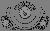
MACHINE PbN.9712 MACH_C0